[最も好ましい] human heart cut in half diagram 130338-Human heart cut in half diagram
14/05/ · The outermost layer of your heart wall is called the epicardium, which is basically a very thin layer of serous membrane The membrane provides lubrication and protection to the outer side of your heart, as you can see in heart diagram labeled Myocardium Right beneath epicardium is another relatively thicker layer called myocardium This muscular middle layer of heart wall contains cardiac muscle tissue Most of the thickness and mass of your heartStart studying Sheep heart cut in half Learn vocabulary, terms, and more with flashcards, games, and other study toolsLearn body parts with felt;
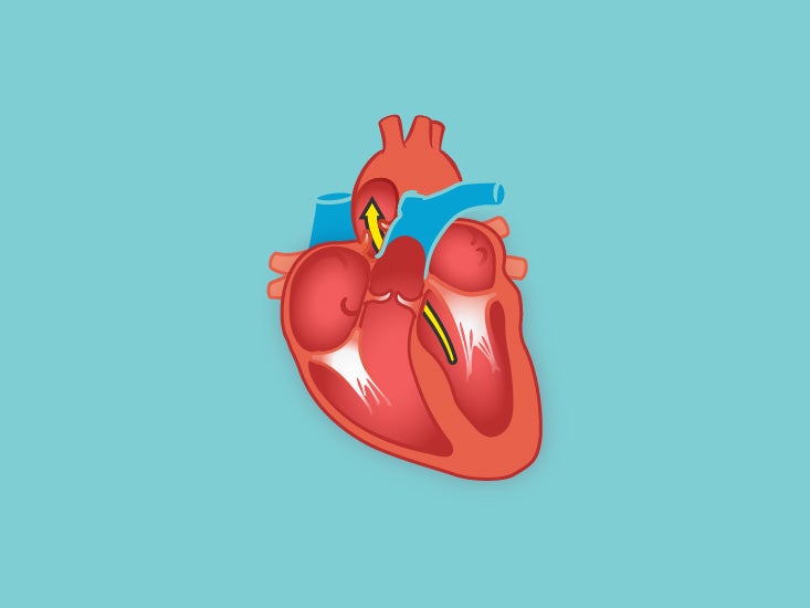
Cross Section Of The Heart Diagram Function Body Maps
Human heart cut in half diagram
Human heart cut in half diagram-The human heart and its functions are truly fascinating The heart, though small in size, performs highly significant functions that sustains human life The human heart resembles the shape of an upsidedown pear, weighing between 715 ounces, and is little larger than the size of the fist It is enclosed in a baglike structure called the pericardium, and is located between the lungs, that isA heart diagram is illustrated in several parts so that it is easily understandable to the learners Students usually have to draw diagrams and learn from pictures given in the text book This diagrammatic representation of human body parts makes it easy for science students to learn about the functionality and working of the organs Below we are going to have a look at various


Cut In Half Face Diagram Quizlet
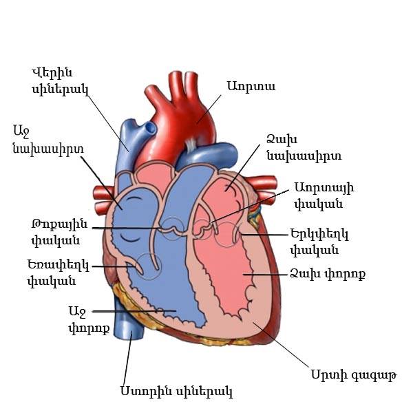
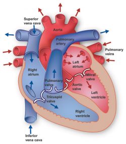
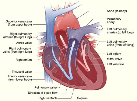
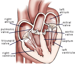
Two ventricles and two atria—both right and left The two left chambers are separated from the two right ones, by a19/02/ · English Diagram of the human heart 1 Superior vena cava 2 4 Mitral valve 5 Aortic valve 6 Left ventricle 7 Right ventricle 8 Left atrium 9 Right atrium 10 Aorta 11 Pulmonary valve 12 Tricuspid valve 13 Inferior vena cava Date , 0702 Source Own work Author Wapcaplet Other versions Derivative works of this file Fontan proceduresvg;27/07/17 · The anterior surface of the human heart faces the sternum, the posterior surface—the base of the cone faces the vertebral column and the inferior or diaphragmatic surface rests on the diaphragm The human heart has got four chambers (Fig 74);
Make a model hand;May 9, Explore Honya Talib's board "Human heart diagram" on See more ideas about Medical knowledge, Nursing study, Nursing school notesCreative Commons "Sharealike" Reviews 49 pbandodkar 2 months ago report 5 thank you
Iv, left ventricle;m, tricuspid valve open ;The structure of the heart If you clench your hand into a fist, this is approximately the same size as your heart It is located in the middle of the chest and slightly towards the leftFeb 24, 21 Explore Juno D Littles's board "Human heart diagram" on See more ideas about heart diagram, human heart diagram, human heart


What S The Difference Between Veins And Arteries Britannica
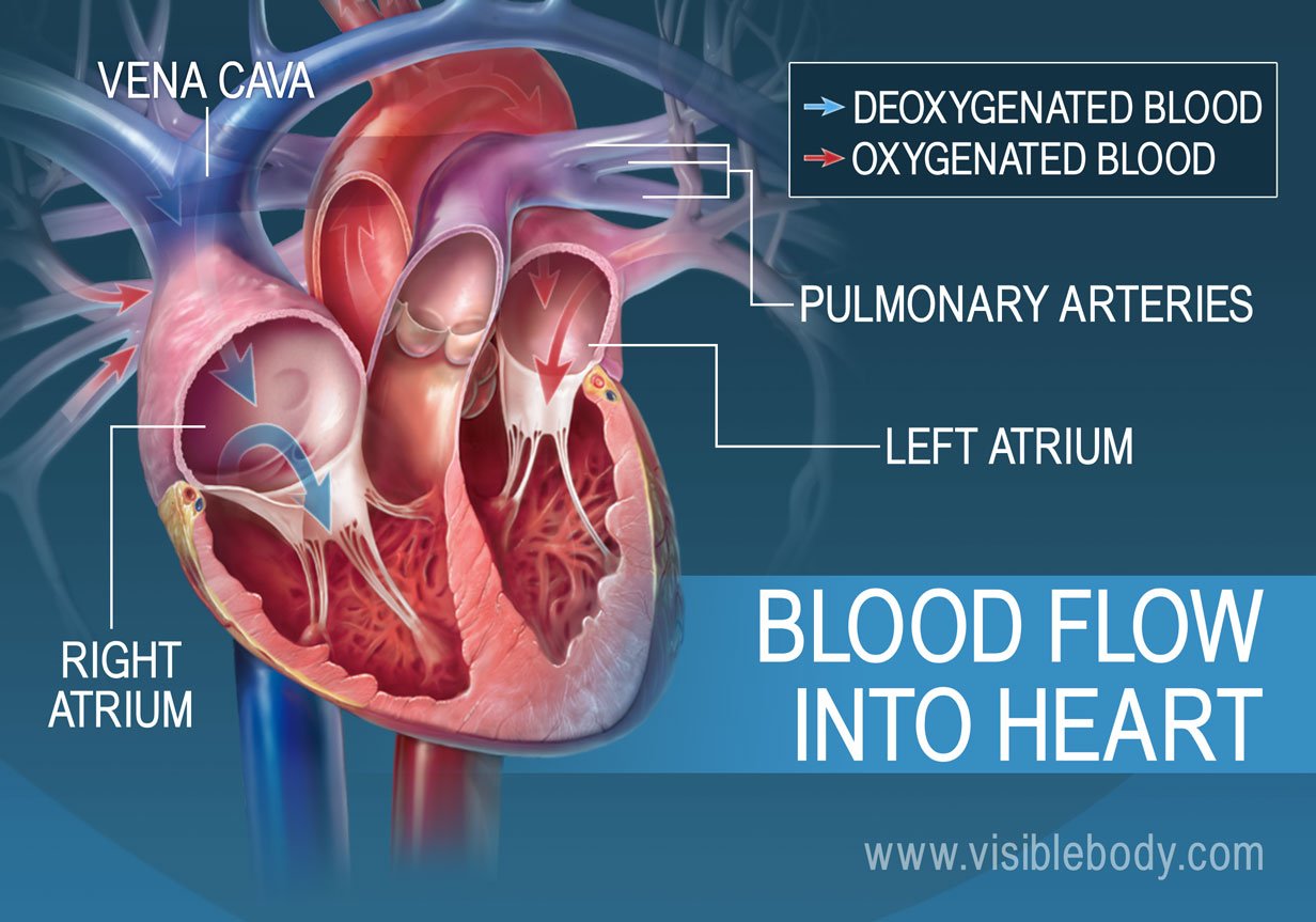


The Heart Circulatory Anatomy
This heart model shows the anatomy of the human heart and is horizontally sectioned at the level of the valve plane The following parts can be removed from heart Esophagus Trachea Superior vena cava Aorta Front more add to cart 3B Smart Anatomy 5 year warranty Heart Model $ 74 Item Full size twopiece normal heart model opens in half to show innerVenison heart is also pretty awesome Cardiac muscle is different from skeletal muscle at the cellular level in a way that results in a meat with little to no 'grain' to it ItHeart cut Noun heartcut (plural heartcuts) A portion of material separated by chromatography that is subjected to heartcutting;
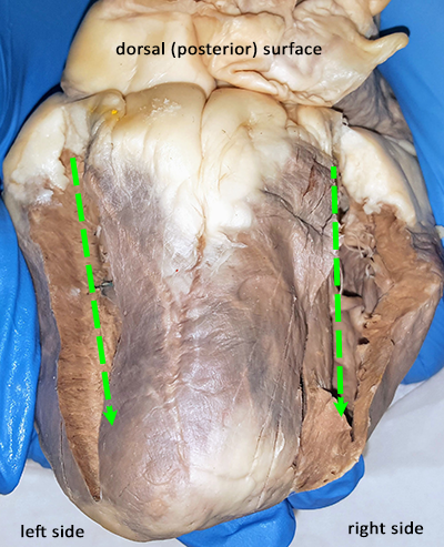


Heart Anatomy Incisions


Cut In Half Face Diagram Quizlet
25/11/14 · Nov 25, 14 This Pin was discovered by Jackie Sweet Discover (and save!) your own Pins on24/04/08 · To draw a human heart, first draw what looks like the lower half of an acorn that's missing its cap This will be the outline of the left and right ventricles Draw a rounded bump on the top left half of the acorn shape, which will be the right atrium Then, sketch a forked tube coming off the top of the rounded bump This will be the superior vena cava, which is what blood entersThe Zygote 3D Human Heart Model Top Cut Model provides a great view of the valves of the heart This model has the atria removed The aorta and the pulmonary trunk are cut just after the aortic valve and the pulmonary valve The 3D heart model top cut is ideal for viewing the valve position in the heart This cut is particularly impressive with the optional animation cycle


Human Heart Labeled Cut In Half Diagram Page 6 Line 17qq Com
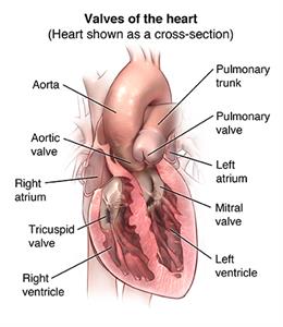


Heart Valve Repair Or Replacement Surgery Johns Hopkins Medicine
Diagram of the circulation of blood as understood after William Harvey's work, showing blood leaving the left ventricle of the heart by the aorta and returning to the heart via the vena cava at the right auricle it has now completed the Greater Circulation It next passes to the right ventricle and out into the Pulmonary Artery and undertakes the Lesser Circulation (Pulmonary Circulation) and returns to the heartYou are basically going to be cutting each side of the heart so that you can look inside (Some dissections will ask you to make a coronal cut where a single cut opens the entire back side of the heart) The heart below is marked to show you where the two incisions should be made Optionally, you may cut the heart in half to expose the chambers My students affectionally call these two variations the "hot dog cut" as pictured above because it looks like a hot dog bun, or the "hamburger cut31 How to Draw A Heart Diagram from Sketch Paint the bottom half of the outline of an acorn until it is tipped to the left Use your pen or pencil to begin drawing the essential part of your heart diagram, which would look like an openended acorn;
/heart_inner_section-577d5c673df78cb62c939314.jpg)


Atria Of The Heart Function
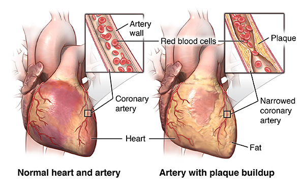


Heart Valve Repair Or Replacement Surgery Johns Hopkins Medicine
13/02/14 · It happens with the heart cycle, which consists of the periodical contraction and relaxation of the atrial and ventricular myocardium (heart muscle tissue) Systole is the period of contraction of the ventricular walls, while the period of ventricular relaxation is known as diastole Note that whenever the atria contract, the ventricles are relaxed and vice versa Let's put into words the heart blood flow diagramN, bicuspid or mitral A^alve closed;p 2CE6YNF from Alamy's library of millions of high resolution stock photos, illustrations and vectorsA lifttheflap body model;
:background_color(FFFFFF):format(jpeg)/images/article/en/heart/yh35Prra0VpHLwLZLrEDA_7dpDZkx6EDgihq981V55Qg_Apex_cordis_02-2.png)


Heart Anatomy Structure Valves Coronary Vessels Kenhub
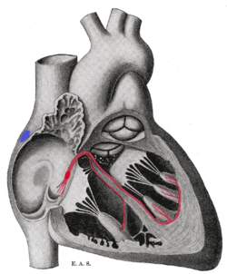


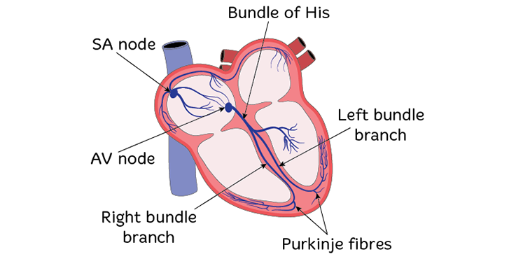
Bundle Of His Wikipedia
Map of the Human Heart Let's get straight to the heart of the matter—the heart's job is to move blood Day and night, the muscles of your heart contract and22/02/18 · This resource from ABPI Schools shows a labelled diagram of the heart Creative Commons "NoDerivatives" Review 4 akudempsey 3 years ago report 4 Report this resourceto let us know if it violates our terms and conditions Our customer service team will review your report and will be in touch £000 Download Save for later £000 Download Save for later LastYou'll find fun activities and experiments for all ages Make your own blood;


Human Heart Diagram In Body Page 6 Line 17qq Com
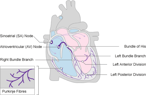


Cardiac Conduction System Normal Function Of The Heart Cardiology Teaching Package Practice Learning Division Of Nursing The University Of Nottingham
The human heart is situated in the middle mediastinum, at the level of thoracic vertebrae T5T8A doublemembraned sac called the pericardium surrounds the heart and attaches to the mediastinum The back surface of the heart lies near the vertebral column, and the front surface sits behind the sternum and rib cartilages The upper part of the heart is the attachment pointCongestive heart failure The heart is either too weak or too stiff to effectively pump blood through the body Shortness of breath and leg swelling are common symptoms Cardiomyopathy A disease of the heart muscle in which the heart is abnormally enlarged, thickened, and/or stiffened As a result, the heart's ability to pump blood is weakenedThis is my second video on the Human heart based on general and previous knowledge which we have been reading for years please go step by step as I am teach


Heart Dissection Estefany S Anatomy And Physiology


Heart Right And Left Atrium Anatomy And Function Kenhub
I, arteries to the lungs;la, left auricle;A brain hemisphere hat;Directions Trace an outline of your child's body on a large piece of butcher paper Using the life size organs on the following pages, place them on your child's body outline Tape or glue them in place along one edge so they can flip up Lift each piece and write the function of the organ underneath the flap


Heart Of The Frog Cut To Half


Heart Anatomy Anatomy And Physiology Ii
22/02/18 · Plain diagram of the heart with labels to add and a cloze exercise on the pathway of blood through the heart Diagram can be coloured as needed and easy to edit in word UPDATED As per request to have an answer sheet Please leave a review to let me know how you get on!Draw the image, and angle it to the left around 1 degrees The critical structure will form the base for ventricles on the left and right;Draw a short wavy line within the original figure, then it extend it up to meet the aortic arch On the other side, extend a long curved line, ending at a small oval Connect this oval to the aortic arch using a curved line that crosses over the end of the arch Human Heart drawing step 4 4
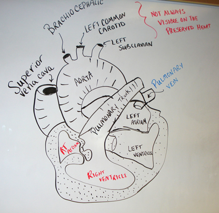


Heart Dissection Walk Through
:background_color(FFFFFF):format(jpeg)/images/article/en/cardiac-cycle/9kXgRmOfS1DdqQ7rymqw_Thorax.png)


Cardiac Cycle Phases Definition Systole And Diastole Kenhub
Explore a virtual body;Fig 76 — Diagram showing thefront half of the heart cut awaya, aorta;18/09/17 · Most heart diagrams show the left atrium and ventricle on the right side of the diagram Imagine the heart in the body of a person facing you The left side of their heart is on their left, but since you are facing them, it is on your right Click for fullsize pdf 1 Identify the right and left sides of the heart Look closely and on one side you will see a diagonal line of blood


Sheep Heart Cut In Half Diagram Quizlet


Diagram Of Human Heart Stock Illustration Download Image Now Istock
How your heart works The human heart works like a pump sending blood around your body to keep you alive It's a muscle, about the size of your fist, in the middle of your chest tilted slightly to the left Each day, your heart beats around 100,000 times This continuously pumps about five litres (eight pints) of blood around your body throughHuman Heart Diagram how to draw in most easy way For class 11th & 12th CBSEhello friend today in this art video i will show you how to draw human heartThe human heart is the most crucial organ of the human body It pumps blood from the heart to different parts of the body and back to the heart The most common heart attack symptoms or warning signs are chest pain, breathlessness, nausea, sweating etc The diagram of heart is beneficial for Class 10 and 12 and is frequently asked in the examinations A detailed explanation of the heart


An Anatomical Review Of The Right Ventricle Sciencedirect


Heart Cut In Half
Adjective heartcut (not comparable) (of a gemstone) Cut in the shape of a heart29/04/ · Within the medulla are several regions of gray matter that process involuntary body functions related to homeostasis The cardiovascular center of the medulla monitors blood pressure and oxygen levels and regulates heart rate to provide sufficient oxygen supplies to the body's tissues The medullary rhythmicity center controls the rate of breathing to provide oxygen to theHeart Diagram Diagram of a heart Human Heart Human Heart Anatomy The human heart consists of the following parts aorta, left atrium, right atrium, left ventricle, right ventricle, veins, arteries and others Heart diagram with labels Human Body Anatomy Diagrams Search Primary menu Skip to primary content Skip to secondary content Bones;


16 The Heart Medicine Libretexts


How Your Heart Works Heart And Circulatory System British Heart Foundation
As you can see, lots of ideas which make discoveringThe pictures below represent a heart that is cut along the horizontal axis The picture on the left shows the plane along which the heart is cut That is, the top of the heart, including the right and left atria (atria is plural for atrium), the pulmonary artery and aorta are removed on the picture on the right (below) It shows the heart as you would look down at it from the front TheInternal structure of human heart shows four chambers viz two atria and two ventricles and couple of blood vessels opening into them The wall of two ventricles are strong and sturdy when compared to atria Before we start, we shall recall the basic proportions of heart and its chambers


Look Good Study Hard Biology Unit 5 2 Transport In Humans
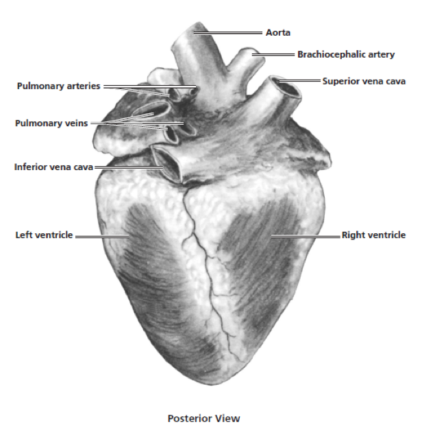


Heart Dissection Carolina Com
Free printable to build a human cell with candy and chocolate;Human heart with vessels, lungs, bronchial tree and cut rib cage Xray effect on black background Creative concept Background of the human heart Vector Illustration eps 10 for your design Heart a Illustration of human heart anatomy Anatomical heart with beams Vector art Vector human heart illustration 4 Views of the human heart from An atlas of human anatomyHome » Uncategorized » Human Heart Diagram Human Heart Diagram The heart is one of the vital organs in the human body It pumps blood throughout the body and supplies nutrients and oxygen to the tissues Oxygenated blood is needed by the body to be active and perform its vital functions The heart beats about 72 times per minute The heart is a muscle that expands and


Brain Cut In Half Diagram Quizlet
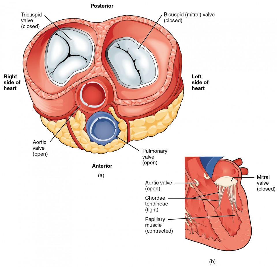


Heart Anatomy Anatomy And Physiology Ii
Find free pictures, photos, diagrams, images and information related to the human body right here at Science Kids Photo name Human Heart Diagram Picture category Human Body Image size 70 KB Dimensions 600 x 600 Photo description This is an excellent human heart diagram which uses different colors to show different parts and also labels aHuman heart diagram highlighting the various anatomical structures The right and the left region of the heart are separated by a wall of muscle called the septum The right ventricle pumps the blood to the lungs for reoxygenation through the pulmonary arteries The right semilunar valves close and prevent the blood from flowing back into the heart Then, the oxygenated blood is30/06/09 · Cardiomyopathy A disease of heart muscle in which the heart is abnormally enlarged, thickened, and/or stiffened As a result, the heart's ability to pump blood is weakened As a result, the heart


16 The Heart Medicine Libretexts
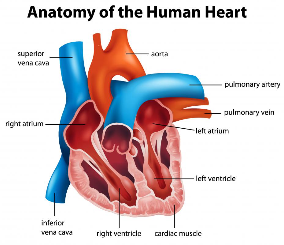


Coronary Sinus Anatomy Anatomy Drawing Diagram
Dissection of the Sheep Heart and Human Heart INTRODUCTION The heart is a coneshaped muscular organ, about the size of your fist It is located in the mediastinum region (central region of the thoracic cavity), between the lungs, and behind the sternum The heart is a hollow organ, containing 4 chambers At least one blood vessel attachesHeart Internal Anatomy Position the heart anterior side up Use your scalpel to cut the heart in half across both atria and ventricles as shown in the figure belowTip Begin at an atrium and cut around the heart to the vessels Open the heart and cut the thick intraventricular septum to complete the separationThe diagram of the skinning pattern is an example of stripstyle skinning, dividing the surface into portions easy to handle Reflect the skin by lifting up and peeling back with one hand, while bringing the knife in as flat to the skin as possible to cut away connective tissue The external genitals present only a small obstacle In the male the penis and scrotum can be pulled away from the


Labeled Human Heart Koibana Info Human Heart Anatomy Anatomy And Physiology Heart Anatomy


Fetal Circulation American Heart Association
29/04/ · Pericardium is a type of serous membrane that produces serous fluid to lubricate the heart and prevent friction between the ever beating heart and its surrounding organs Besides lubrication, the pericardium serves to hold the heart in position and maintain a hollow space for the heart to expand into when it is full The pericardium has 2 layers—a visceral layer that covers the outside of the heartListen to your heart with a cardboard tube stethoscope;
:max_bytes(150000):strip_icc()/cardiac_cycle-597a5d8168e1a200115e5937.jpg)


The Anatomy Of The Heart Its Structures And Functions


Cool Anatomy Slides Album On Imgur


Human Heart Labeled Cut In Half Diagram Page 1 Line 17qq Com


Human Heart Diagram And Anatomy Of The Heart


Unit Four Lsa
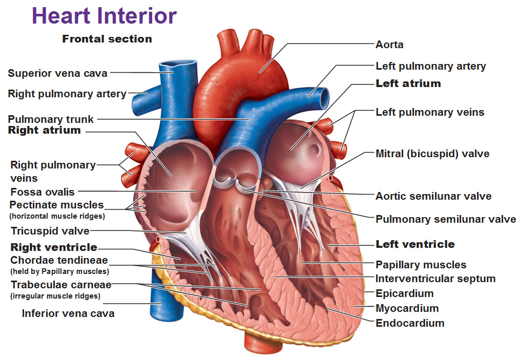


Blood Flow Of The Heart


Circulatory Systems In Animals Transport Systems In Animals Siyavula


Blood Flow Of The Heart


Science For Kids Cardiovascular System


Heart Information Center Heart Anatomy Texas Heart Institute


Heart Transplantation Wikipedia


Human Heart Labeled Cut In Half Diagram Page 7 Line 17qq Com


Pin On Anatomia
:background_color(FFFFFF):format(jpeg)/images/article/en/conducting-system-of-the-heart/AQP7Vp65mF6GLrKpnbUw_Heart_anterior_view.png)


Conduction System Of The Heart Parts And Functions Kenhub


Cross Section Of The Heart Diagram Function Body Maps


Diagram Of Human Heart Stock Illustration Download Image Now Istock


Pin On School Stuff


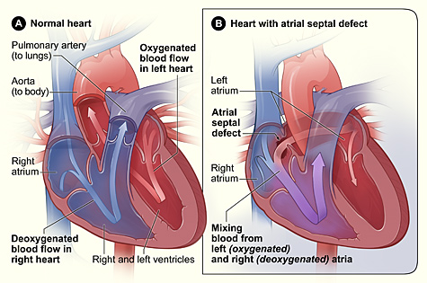
Adult Cardiothoracic Surgery Ventricular Septal Defect


Blood Flow Of The Human Heart Free Vector On Freepik


How The Heart Works Heart Foundation


16 The Heart Medicine Libretexts
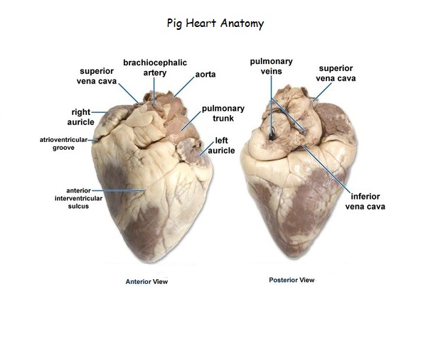


Heart Dissection


V Angiology 4b The Heart Gray Henry 1918 Anatomy Of The Human Body


Thoracic Cavity An Overview Sciencedirect Topics


Free Printable Heart Diagram For Kids Labeled And Unlabeled
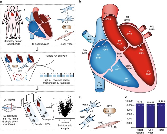


Region And Cell Type Resolved Quantitative Proteomic Map Of The Human Heart Nature Communications


How To Draw Heart Diagram In Exams Youtube


Anatomical Planes Of Body What Are They Types Position In Body


Complete Heart 3d4medical


Free Printable Heart Diagram For Kids Labeled And Unlabeled
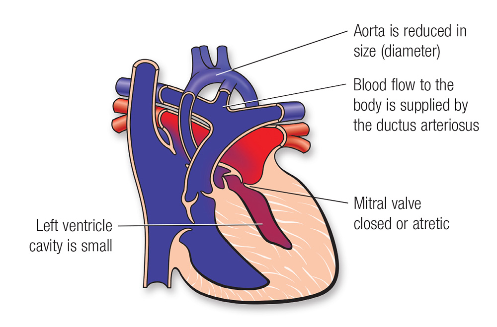


Single Ventricle Defects American Heart Association
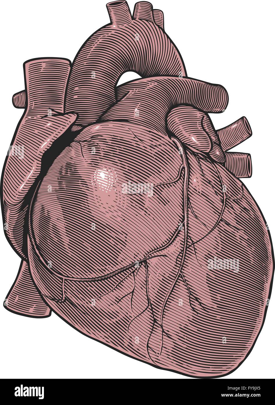


Human Heart High Resolution Stock Photography And Images Alamy
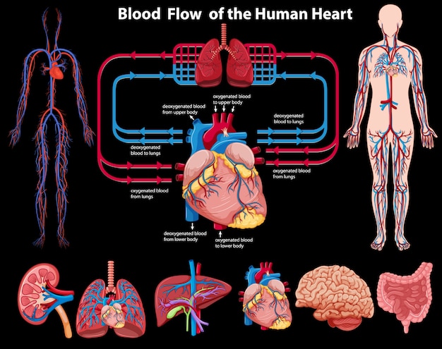


Blood Flow Of The Human Heart Free Vector On Freepik


40 3a Structures Of The Heart Biology Libretexts


The Proposal Danielle Heyder S Blog For Alfred University Foundations Course
/heart_posterior_aorta-57f663543df78c690f0885c1.jpg)


Anatomy Of The Heart Aorta
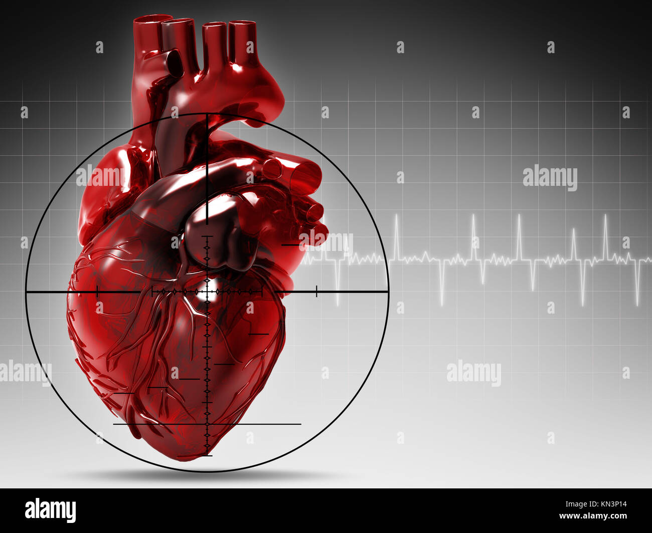


Human Heart High Resolution Stock Photography And Images Alamy


109 Single Coronary Type R2a Dr Buchanan S Cardiology Library Vin


Heart And Circulatory System
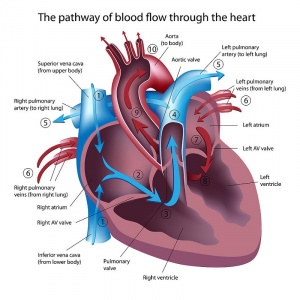


Pharmacological Management Of Congestive Heart Failure Physiopedia


Draw A Diagram To Show The Internal Structure Of Human Heart Label 6 Parts In All Including Atleat Brainly In
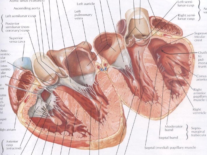


Heart


Meet The Heart Video Human Body Systems Khan Academy


Heart Dissection Carolina Com


File Diagram Of The Human Heart Cropped Svg Wikimedia Commons


Heart Anatomy Anatomy And Physiology Ii


How The Heart Works Diagram Anatomy Blood Flow


Heart Real Human Heart Anatomy
:background_color(FFFFFF):format(jpeg)/images/library/8671/heart-in-situ_english.jpg)


Circulatory System Structure Function Parts Diseases Kenhub


Blood Flow Of The Human Heart Free Vector On Freepik


Seer Training Structure Of The Heart
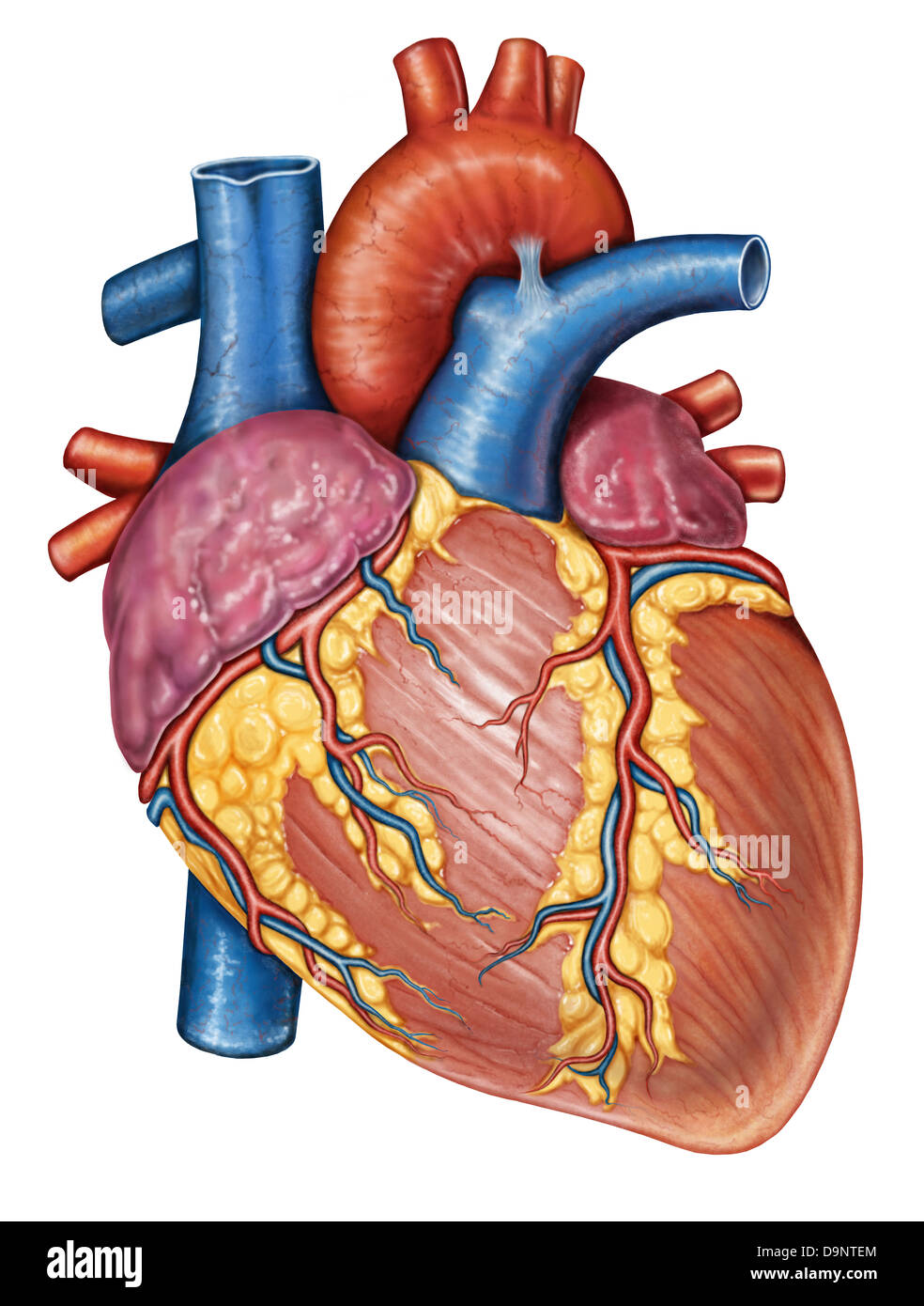


Human Heart High Resolution Stock Photography And Images Alamy


Anatomical Planes Of Body What Are They Types Position In Body


Real Human Heart Cut In Half Page 1 Line 17qq Com


Human Heart Stock Illustration Illustration Of Diagram
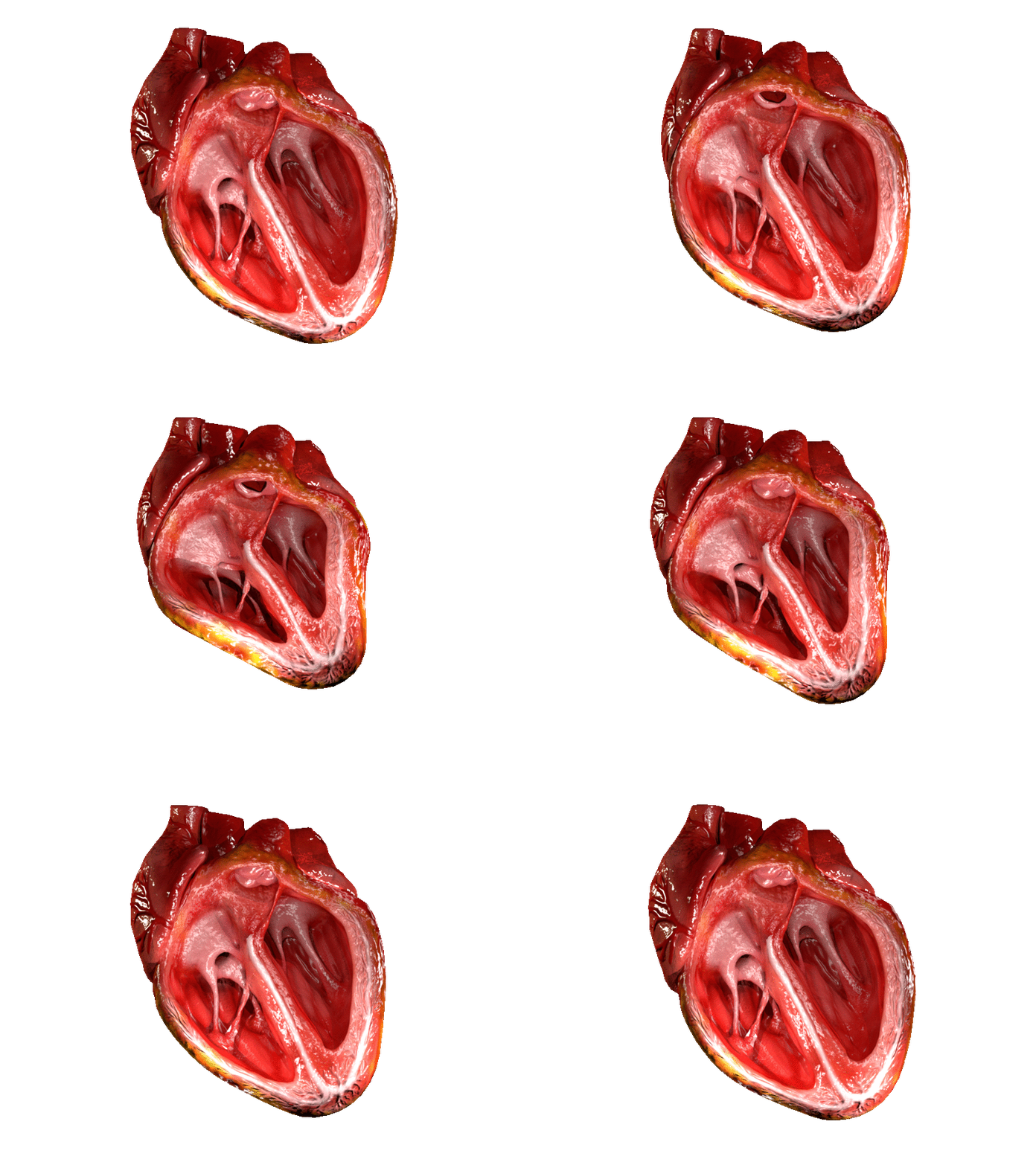


Heart Valve Wikipedia


Circulatory Systems In Animals Transport Systems In Animals Siyavula


16 The Heart Medicine Libretexts
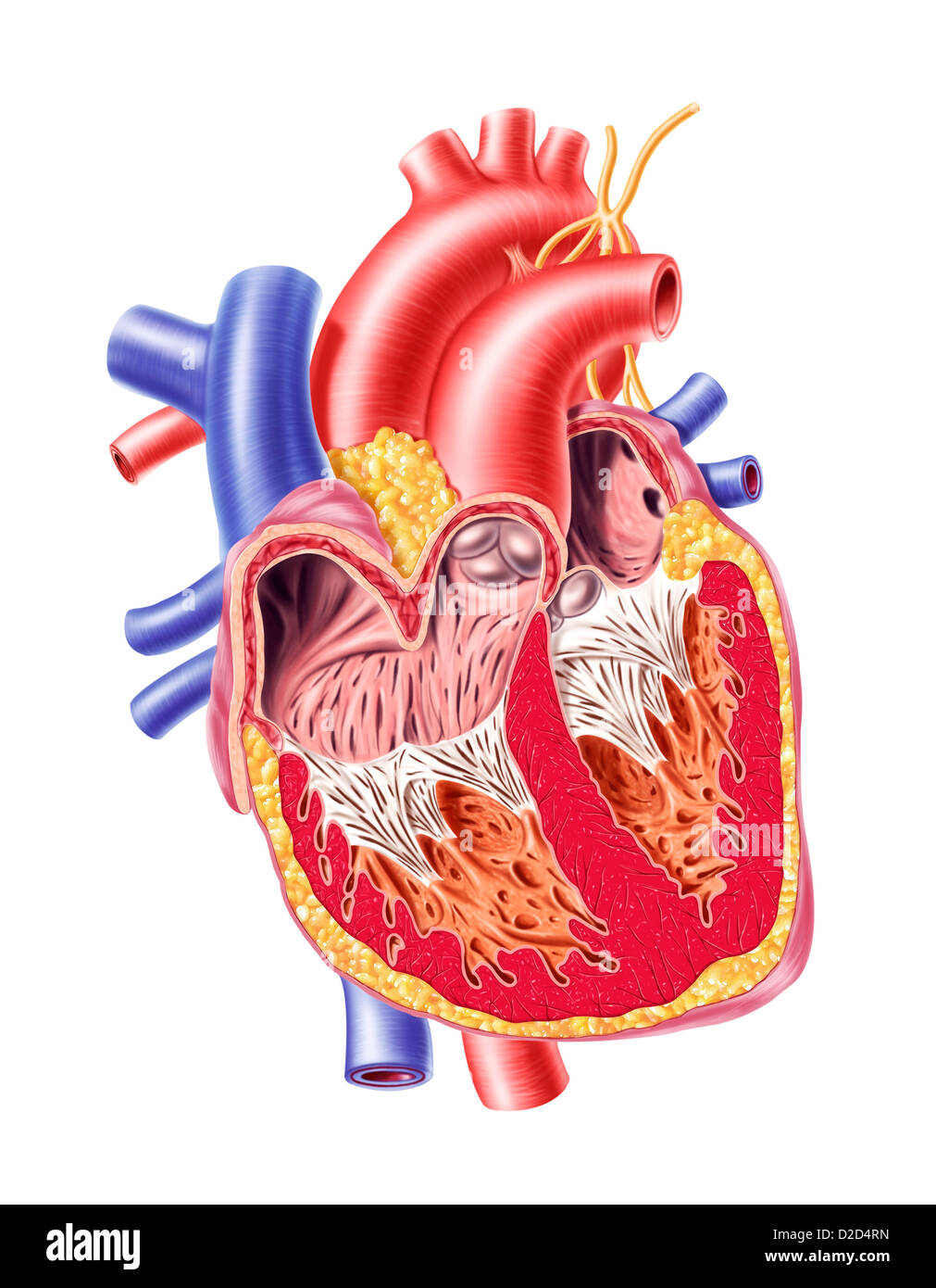


Human Heart High Resolution Stock Photography And Images Alamy


Human Heart Labeled Cut In Half Diagram Page 1 Line 17qq Com
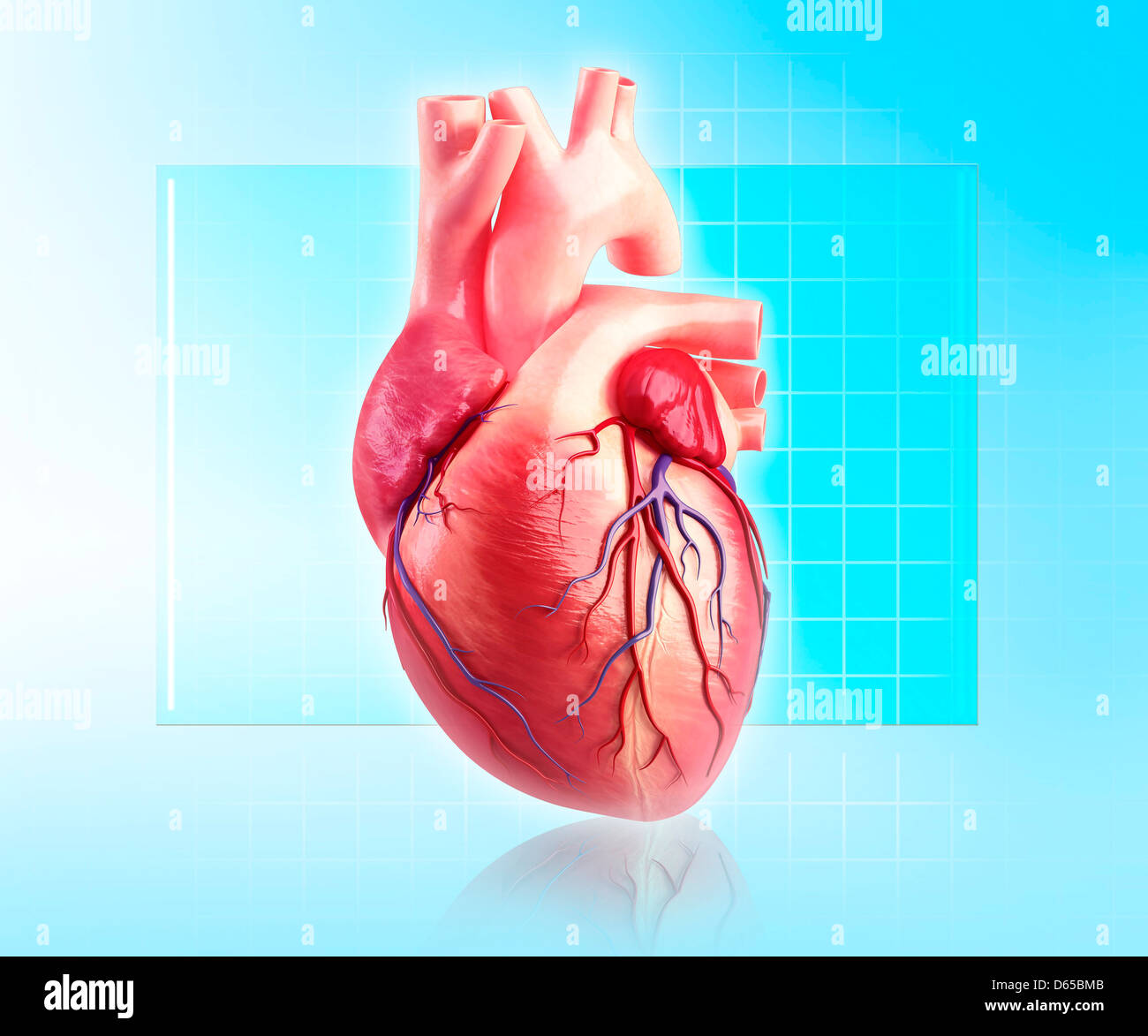


Human Heart High Resolution Stock Photography And Images Alamy


Congenital Heart Conditions


How The Heart Works Heart Foundation


Human Eye Definition Structure Function Britannica


Adult Cardiothoracic Surgery Ventricular Septal Defect


コメント
コメントを投稿